
CT Dental Scans
The iCAT CT Dental Scan allows us to "pre-plan" many procedures, saving time, answering previous unanswerable questions and minimizing risk and guesswork before surgery. With the "Teeth in an Hour" protocol, the patient can leave the chair with teeth in place after a short and non-traumatic surgery and experience a minimum number of post-operative reactions. This new modality provides enhanced patient satisfaction and simplifies the process of the treatment team. Read our CT Dental Scan FAQ below.
We are now able to offer our patients a new procedure called a CT Dental Scan to make surgery even safer and more predictable. See below for answers to frequently asked questions regarding this breakthrough technology: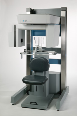
- What is a CT Dental Scan?
- How does an CT Dental Scan differ from conventional X-rays?
- So why do I need such a specialized X-ray?
- Do I need to do anything special? And what will happen to me during the CT scan?
- What about the radiation aspects?
- So where do I go?
What is a CT Dental Scan?
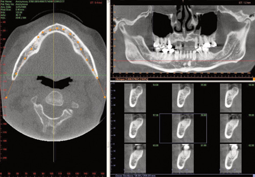
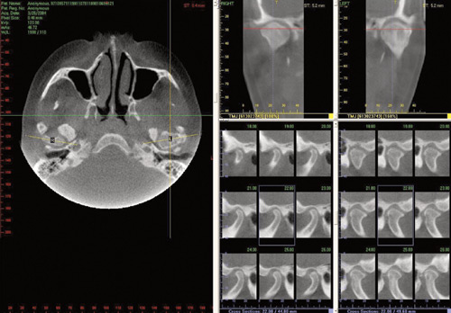
CT Dental Scan uses advanced computer programs to analyze an X-ray study called a CT scan. By providing detailed two-dimensional and three-dimensional images, CT Dental Scans enable your dentist to select the best location for your implants and plan the details of your surgery with pinpoint accuracy, well before the operation. The main benefit is less guesswork.
How does a CT Dental Scan differ from conventional X-rays?
Routine dental x-rays are two-dimensional; they only show the location of your teeth and the height of the bone. These x-rays are often distorted and cannot depict the 3-Dimensional thickness of your jawbone.
A CT Dental Scan, on the other hand, is distortion free. It illustrates the actual make-up of the bone and provides three-dimensional and cross-sectional views of your jaws. The life-sized images allow us to accurately measure the amount of bone that is available for implants.
So why do I need such a specialized X-ray?
CT Dental Scans help us to determine if you are a good candidate for implant surgery or one of our other services. This can save you unnecessary operations, time and money. Because the 3-D image tells us a lot more than conventional x-rays, it helps us better prepare for your surgery. A CT Dental Scan will show us the exact location of anatomical structures, the contours of the jaw bone, and the best sites for your implants before going into surgery.
This means that we can virtually eliminate surprises when we perform your surgery. That means less operating time and fewer complications for you.
Do I need to do anything special? And what will happen to me during the CT scan?
You do not need to prepare for the CT scan. You may be asked to remove any jewelry from your head and neck so that it does not interfere with the study. Once in the examination room, all that you have to do is to rest face up on the CT scanner bed. Your head will be comfortably cushioned on a padded cradle, and a velcro strap will hold your head so that it remains still.
Keeping still for only eight seconds is very important so that clear pictures are obtained. The scan will be done before you know it.
What about the radiation aspects?
The radiation dose received varies from patient to patient but is typically in the range of 0.1 to 0.5 mSv. To put this in perspective, everyone in the country receives about 2.2 mSv annually from natural background radiation, so a CT Dental Scan corresponds to less than an additional three months of background radiation. The background radiation is less for our Cone-Beam compared to other Medical based Spiral CT Scanners.
So where do I go?
Right in our office! Just contact us and make an appointment that is convenient for you.
CT Dental Scan Detailed Specifications
- High Resolution Scan produces images at .2mm voxel size.
- Fast Scan Time (8.9 seconds) minimizes radiation and reduces chances of patient movement during scan - quicker and easier image acquisition.
- Adjustable Beam Collimation allows full height and targeted field of view scans, providing the ability to further minimize patient radiation.
- Comfort Design positions patient in an open environment seated position - allows patient comfort and capture of natural orientation of anatomy.
- Fast Reconstruction Time provides the most complete views of all oral and maxillofacial structure in real-time - produces three-dimensional images typically under one minute.
- Easy To Operate point-and-click remote makes the i-CAT easy to use.
- DICOM 3 Compatible data makes it easy to share and integrate with third party volume data software companies.
- 12 bit Grayscale quality allows more shades of gray to increase contrast for easier viewing.
- Report Printout function provides full-color reports with sophisticated clarity and precision.
- Priced Affordably.
- X-ray Source: High Frequency, Constant Potential, fixed anode 120 kVp, 3-8 mA (Pulse mode).
- X-ray Beam: Cone
- Focal Spot: 0.5mm
- Image Detector: Amorphous Silicon Flat Panel, 20 cm x 25 cm
- Gray Scale: 12 bit
- Voxel Size: 0.4 mm (typical), 0.2 mm (minimum)
- Image Acquisition: Single 360 degree rotation
- Scan Time: 20 seconds standard (options of 10, 20, 40)
- Patient Position: Seated
- Scan Dimensions: 17 cm (diameter) x 13 cm (height)
- Primary Reconstruction: 1.5 minutes for standard 20 second scan
- Secondary Reconstruction: Real Time