
Dental Implants are highly predictable and have the highest level of long-term success rates found in dentistry. Implants are used to replace single or multiple missing teeth with crowns as well as secure loose appliances/ replace a failing set of teeth with removable or fixed/ screwed-in options. According to a 15-year-study and report, titanium implants have greater than a 90% success rate and are still present and in function after that period of time. Dental Implants can be made from Titanium or Zirconium ceramic materials. Titanium is well accepted by the human body and can integrate with the alveolar bone while Zirconium Ceramic implants are just now being reviewed as an alternative to a metal based implant. Only titanium implants are placed at this office currently.
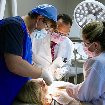
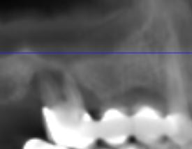
Missing or clinically hopeless teeth create a difficult time with eating (masticating) food and can also be considered unsightly. A dental implant can be placed as long as the amount and quality of bone provides adequate support for the fixture. Having a Dental CT scan taken and reviewed prior to implant placement can help determine if the amount and quality of bone is adequate for an implant. As part of our planning process, a Dental CT scan will be taken at Engle Dentistry prior to a final treatment plan being presented. With this single scan, Dr. Engle can identify if any pre-surgical infections exist or if the anatomy of the bone requires any additional bone growing techniques prior to or during the placement of the dental fixture. Understanding the amount and quality of bone prior to starting any definitive treatment will maximize your level of implant success.
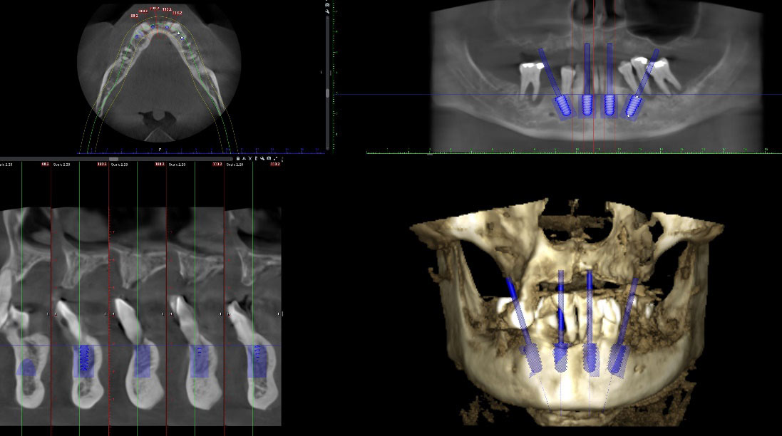
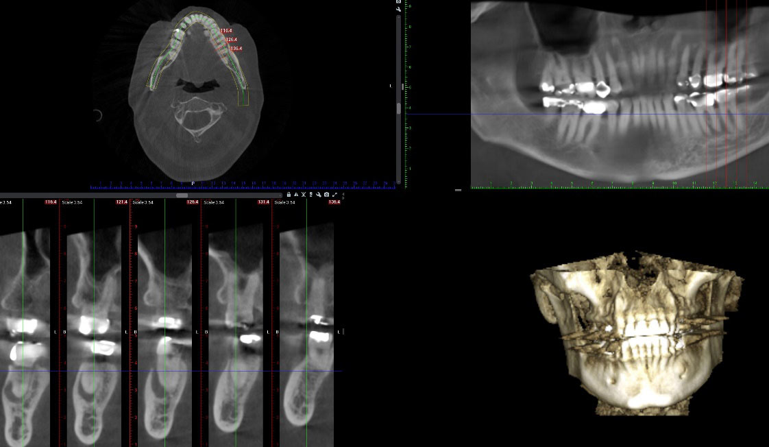
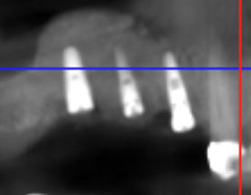
Dental CT Radiographs: 3D Radiographs Benefits include:
- An effective tool in the surgical planning for dental implants and removal of wisdom teeth
- Evaluation of the Sinuses as well as position of important anatomy including nerve canals
- Detecting and diagnosis of jaw tumors, pathology and anomalies
- Provides accurate details of both the bone and soft tissue
Implants can be used to replace single tooth, more than one missing teeth or even a full arch of teeth. Wearing a removable appliance is very challenging to most patients. Partial dentures can have unsightly wires while full dentures are difficult to eat with and sometimes a challenge to keep secured in the mouth. With the advances in dental materials, a full arch of ceramic (Zirconium) or Nanoceramic (Crystal Ultra®) appliances is possible and can be fabricated with enough strength to avoid chipping and breaking of the appliance. These alternative materials avoid staining, are very strong to chewing forces and are extremely aesthetic compared to Acrylic appliances.
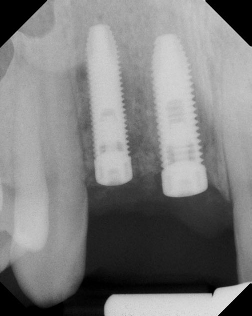
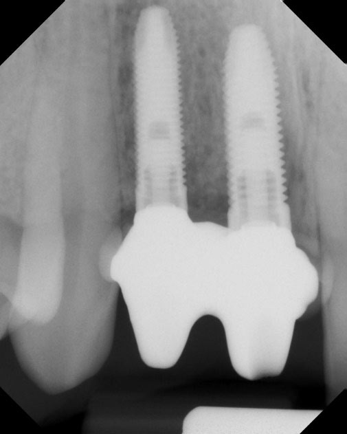
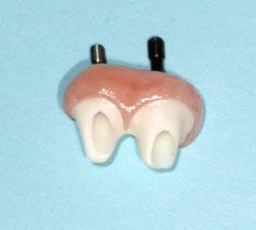
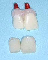
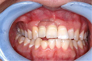
Case #1: Two Front Teeth Knocked Out From a Fall
Unfortunately, when the teeth were forced out, so did a huge piece of his bone. Even after the repair with bone grafting, we knew that we were not going to get a 100% aesthetic result without the use of pink porcelain. He was presented with two treatment options: Titanium Dental Implants vs. Cemented Fixed Bridge.
He elected to have two dental implants placed.
Two dental implants were placed to the best position based upon what was available to work with. He was given a removable appliance to wear until the implants could heal. After four months, final impressions were taken and sent to a ceramic lab. A two-implant bridge was custom fabricated to fill the remaining bone defect as well as to provide for optimal aesthetics. Working with the right ceramist, this aesthetic result was possible.
Case #2: Failing Bridge with Infected Molar
Inadequate bone was available for dental implants with bone grafting needed to both the ridge and the sinus area. This procedure was staged between two different phases.
Phase #1 included the surgical removal of the infected tooth with bone grafting to both the ridge and infected tooth areas.
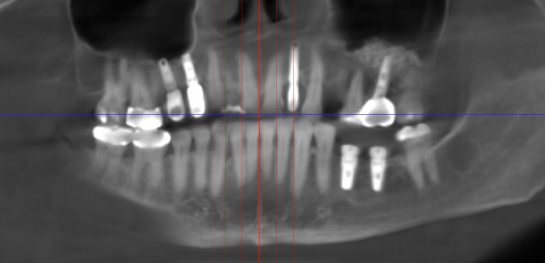
Phase #2 After 3 months of healing, the placement of the three(3) dental implants with subantral augmentation (Sinus Lift). An additional 5 months of healing was required prior to the fabrication of the final implants crowns.
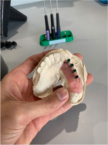
Case #3: Implants Help Secure Loose Appliances Using Little Nylon Snaps (Locators)
Patients that wear dentures get frustrated because they are unable to eat steak and are forced to eat softer foods because the appliances just flop around. It is a more economical solution to have a removable implant secured appliance compared to a fixed one. When considering removable, the lower arch requires two, four or sometimes six implant fixtures. Compared to the upper arch which requires at least five implants to get rid of the palate being covered with acrylic.
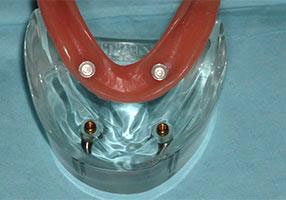
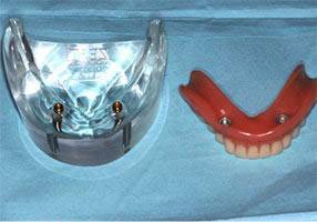
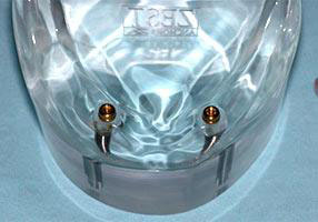
Small nylon snaps hold the appliance secure. These attachments do come in different retentive strengths. The clear ones highlighted on the sample model have the best retention and are the strongest available.